Fig. 1.
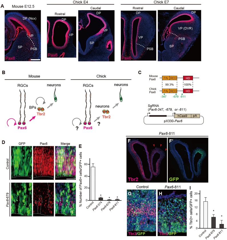
Targeted deletion of the endogenous Pax6 gene in the developing chick pallium. (A) Expression patterns of Pax6 protein in mouse (E12.5) or chick (E4 and E7) telencephalon. DP, dorsal pallium; Ncx, neocortex; PSB, pallium-subpallium boundary; SP, subpallium; VP, ventral pallium; DVR, dorsal ventricular ridge. (B) Interspecies differences in mouse and chick pallial neurogenesis. BPs, basal progenitors; RGCs, radial glial cells. (C) (Top) Protein structures of mouse and chick Pax6 showing the percentage identity of domains. The paired domain consists of PAI and RED subdomains; HD, homeodomain. The location of the three sgRNA target sites is indicated. (Bottom) The pX330-Pax6 vector for simultaneous expression of sgRNA and Cas9. (D,E) Electroporation of pX330-based vectors into the developing chick pallium. The number of Pax6-positive cells among GFP-positive cells is significantly decreased by electroporation with pX330-Pax6 vectors (E). Welch's t-test, *P<0.05. Error bars indicate s.e.m. n=4. (F-I) Decreased number of Tbr2-positive cells after electroporation of pX330-Pax6. Arrowheads indicate the reduction of Tbr2 expression in the electroporated region. There was no significant difference in the proportion of Tbr2-positive cells between Pax6-679 and -811, indicating that the two sgRNAs were equally efficient in targeting chick Pax6. Error bars indicate s.e.m. n=4 for control, n=3 for Pax6-679 and -871 samples. Two-tailed Student's t-test, *P<0.05. Scale bars: 200 µm in A; 25 µm in D.

