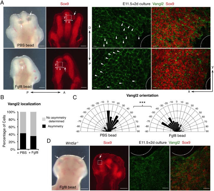
Fig. 2.
Fgf signaling is sufficient to regulate PCP in a Wnt5a-dependent manner. (A) Representative morphological and immunofluorescence images of cultured distal limbs. Beads soaked in PBS or Fgf8 recombinant protein were inserted into E11.5 mouse embryonic forelimbs, which were then cultured for 2 days. The boxed regions lateral to the beads (arrows) were scanned by confocal microscope and representative images are shown in the middle (Vangl2, green) and right-hand panels (Vangl2/Sox9, green/red). The beads are outlined. Arrowheads point to some of the asymmetrically localized Vangl2. P-D and A-P axes are indicated. x- and y-axes of the images are defined as shown in the left-hand panels. (B) The percentage of cells with discernible Vangl2 asymmetric localization in digits implanted with PBS- or Fgf8-soaked beads. (C) Schematics summarizing the quantification of orientation angles of Vangl2. x-axis, angle of orientation (−90° to 90°); y-axis, percentage of cells at angle x. Kolmogorov–Smirnov test, ***P=0.0008. (B,C) PBS beads-implanted limbs N=4, analyzed cells n=711; Fgf8 beads-implanted limbs N=5, analyzed cells n=678. (D) Loss of Vangl2 asymmetric localization in Wnt5a−/− limbs cannot be rescued by Fgf8-soaked beads after 2 days of culture. Boxed region close to the bead (arrow) is shown in the middle (Vangl2, green) and right-hand panels (Vangl2/Sox9, green/red) at higher magnification. The bead is outlined. No Vangl2 asymmetric localization was observed. Cultured Wnt5a−/− limbs: N=4. Scale bars: 200 μm (left-hand panels of A and D); 20 μm (right-hand panels of A and D).

