Fig. 7.
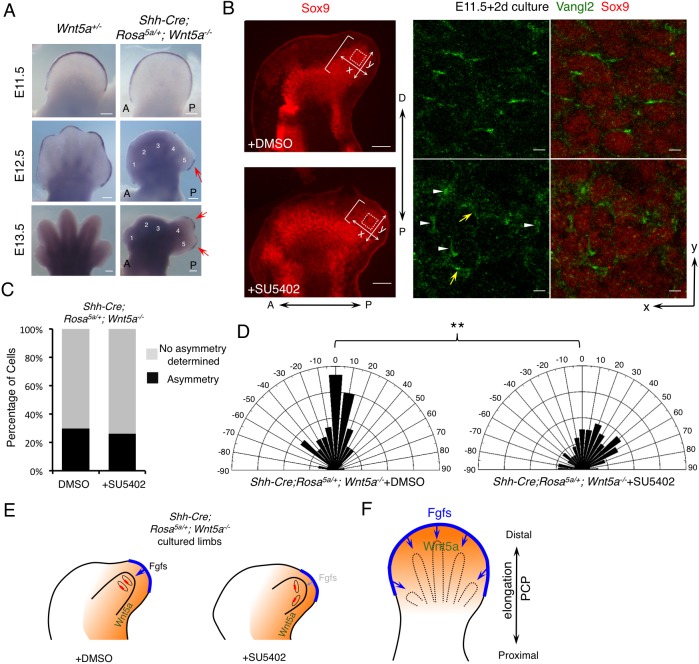
Inhibition of distal Fgf signaling renders cells more responsive to oriented Wnt5a signaling. (A) Whole-mount in situ hybridization of Fgf8 in forelimbs of the embryos with the indicated genotypes. Fgf8 expression was restored (red arrows) in the AER overlying the posterior digits of Shh-Cre; Rosa5a/+;Wnt5a−/− embryos. A, anterior; P, posterior. Digits 1-5 are labeled in the mutant. (B) Treatment with Fgf receptor inhibitor (SU5402) changed the direction of Vangl2 asymmetric localization in cultured Shh-Cre;Rosa5a/+;Wnt5a−/− embryonic forelimbs (E11.5+2-day culture). The brackets indicate the length of digit outgrowth. Part of the boxed regions (dotted squares) is shown in the middle (Vangl2, green) and right-hand (Vangl2/Sox9, green/red) panels at higher magnification. White arrowheads point to 90° oriented Vangl2. Yellow arrows point to Vangl2 with other orientations. P-D and A-P axes are indicated. x- and y-axes of the boxed regions are defined as shown in the left-hand panel. (C) The percentage of cells with discernible Vangl2 asymmetric localization in distal chondrocytes of Shh-Cre;Rosa5a/+;Wnt5a−/− limbs after DMSO or SU5402 treatment. (D) Schematics summarizing the quantification of orientation angles of Vangl2 in each group. x-axis, angle of orientation (−90° to 90°); y-axis, percentage of cells at angle x. Kolmogorov–Smirnov test, **P=0.0027. (C,D) DMSO-treated limbs N=6, counted cells n=414; SU5402-treated limbs N=6, counted cells n=406. (E) Schematics summarizing the results shown in B-D. The red boxes represent Vangl2 asymmetric localization and the blue arrows represent Fgf signaling. The orange backgrounds represent Wnt5a signaling. The light blue arrow in the right-hand panel indicates weaker Fgf signaling. (F) Schematic of the proposed model: PCP establishment is coordinately regulated by both Wnt5a and Fgfs signaling. Orange represents graded Wnt5a expression; blue, Fgf signaling from the AER. Scale bars: 200 μm (A and left-hand panels of B); 10 μm (right-hand panels of B).

