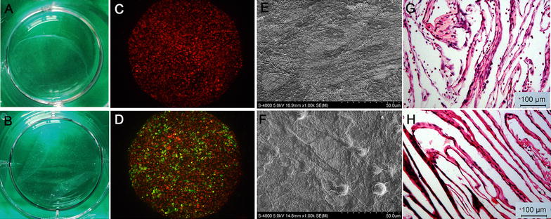
Fig. 1.

Structure of cell sheets. Macroscopic images of cell sheets of the BMSC group (A) and +EPC group (B) detached from culture dishes. Representative microscopic view of cell sheet morphology of the BMSC group (C) and +EPC group (D); BMSCs were stained with Dil (red) and EPCs were stained with DiO (green). Representative SEM images of cell sheets of BMSC group (E) and +EPC group (F). H&E staining images (×100) of cell sheet of BMSC group (G) and +EPC group (H)
