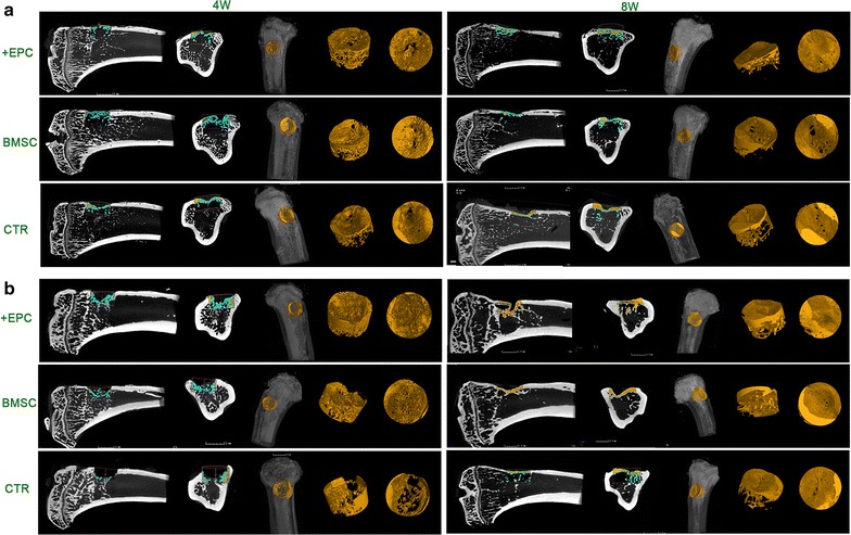
Fig. 5.

Micro-CT evaluation of newly formed bone. 2D and 3D images of the bone formed in the defect area of non-irradiated (a) and irradiated (b) rats at 4 and 8 weeks. Bone structure within the ROI is shown in orange in the 3D reconstructed images. The lateral and coronal views of the reconstructed defect area are shown
