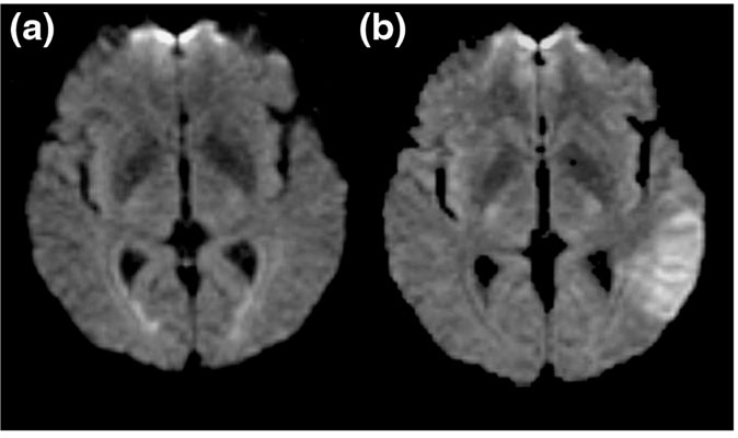
Figure 1.

The diffusion weighted imaging (DWI) of 3 h stroke. (a) The fluid-attenuated inversion recovery image without diffusion weighting shows no acute lesion. (b) DWI demonstrates the acute lesion as a region of hyperintensity (brightness) in the left temporal lobe.
