Fig. 1.
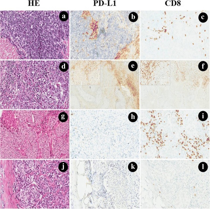
Hemotoxylin and eosin (H&E), PD-L1 and CD8 stains, each performed on histologic sections of small cell lung cancer (a-c), large cell neuroendocrine carcinoma (d-f), atypical carcinoid (g-i) and typical carcinoid (j-l). a Small cell lung cancer was showing a scant cytoplasm, fine nuclear chromatin, absent or inconspicuous nucleoli, extensive necrosis. b PD-L1 was moderately expressed on the membrane of stromal immune cells in the desmoplastic stroma between clusters of tumor cells. c CD8+ TILs were observed in the stroma, while the intratumoral pattern of CD8 expression was not common. d Large cell neuroendocrine carcinoma with prominent nucleoli and abundant eosinophilic cytoplasm, necrosis was not shown. e, f PD-L1 was positively expressed on the membrane and cytoplasm of the immune cells, and a large number of CD8+ TILs could also be observed at the borderline (the 200× magnification for PD-L1 and CD8 was shown in the upper left, respectively). g Atypical carcinoid with vascularized stroma, focal necrosis and 6 mitosis/2 mm2. h, i PD-L1 was negative expression either in tumor cells or stromal cells, while CD8+ TILs were exhibited in the interface of tumor and stroma. j Typical carcinoid with organoid growth pattern with intervening vascular stroma. k, l No PD-L1 can be detected, and only several CD8+ TILs could be found in the stroma. (The original magnification of e-f was 100×, magnification for remaining cases were 200×)

