Fig. 3.

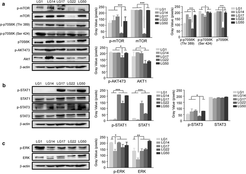
Activated signal transduction pathways in the established lung cancer PDXs. a AKT-mTOR axis pathway was activated in the different PDXs (LG1, LG14, LG17, LG22 and LG50). Protein levels of p-mTOR and mTOR, p-p70S6K (Thr389), p-p70S6K (Ser424) and p70S6K, p-AKT473 and AKT1 were visualized by Western blotting. β-actin was used to verify equal protein loading. b STAT1 and STAT3 pathway were activated in the different PDXs (LG1, LG14, LG17, LG22 and LG50). Protein levels of p-STAT1 and STAT1, p-STAT3 and STAT3 were visualized by Western blotting. β-actin was used to verify equal protein loading. c ERK pathway were activated in the different PDXs (LG1, LG14, LG17, LG22 and LG50). Protein levels of p-ERK and ERK was visualized by Western blotting. β-actin was used to verify equal protein loading. Each experiment was repeated three times. According to the results of Western blotting, we used image J to determine the gray value of each stripe, made histograms by the gray value, the asterisks (*,**, ***) indicate a significant (p < 0.05, 0.01, p < 0.001, respectively)
