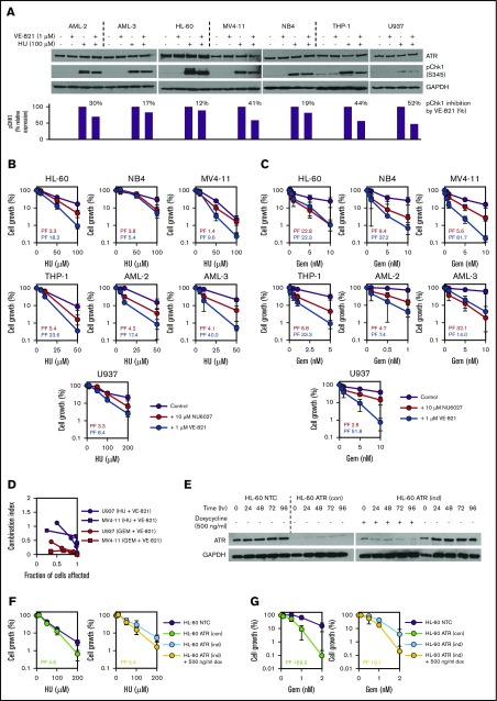
Figure 1.
ATR inhibition using small molecule inhibitors or shRNA-mediated gene knockdown potentiates the inhibitory effects of hydroxyurea (HU) and gemcitabine (Gem) in AML cell lines. (A) To confirm target engagement, AML cell lines were exposed to 100 µM HU (or vehicle control) for 1 hour in the presence of 1 µM VE-821 (or DMSO control), and western blotting was performed for ATR and pCHK1 (Ser345). Expression of pCHK1 is expressed relative to the HU-only lane for each cell line. (B-C) AML cell lines were treated with (B) HU or (C) Gem either alone (purple circles), in combination with 10 µM NU6027 (red circles), or in combination with 1 µM VE-821 (blue circles), and cell density (relative to respective vehicle controls) was determined after 96 hours. PFs were calculated for both ATR inhibitors (NU6027, red text; VE-821, blue text) at the highest dose of HU or Gem. (D) U937 and MV4-11 AML cells were treated with escalating does of VE-821 and/or drug (Gem or HU). CI values <1 indicate synergy between drug combinations. (E) Western blotting for ATR was performed at 24-hour intervals to confirm shRNA-mediated knockdown in HL-60 ATR (con) cells with constitutive ATR knockdown (left blot) and HL-60 ATR (ind) cells with doxycycline-induced ATR knockdown (right blot). (F-G) HL-60 ATR (con) cells (green circles, left chart) and HL-60 ATR (ind) cells (yellow circles, right chart) and their respective controls were treated with (F) HU or (G) Gem, and cell growth and PFs were determined as above. For all western blots, glyceraldehyde-3-phosphate dehydrogenase (GAPDH) was used as a loading control, and for all potentiation assays, data represent the mean ± standard deviation (SD) of 3 independent experiments. See supplemental Figures 2, 3, and 5A. dox, doxycycline; NTC, non-target control.

