Fig. 1.
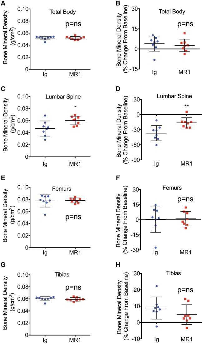
BMD is significantly increased in the lumbar spine following MR1 treatment
Mice (female C57BL6) were administered Ig (control) or MR1 for 6 months, beginning at 5 months of age. BMD was quantified by DEXA at baseline and at 11 months of age and data are presented as BMD (g/cm2), as well as BMD (% change from baseline) for total body BMD (A and B), lumbar spine (C and D), femur (E and F) and tibia (G and H). Mean (s.d.). *P < 0.05; **P < 0.01; by Student’s t-test. n = 8 Ig and 8 MR1 mice/group. DEXA: dual energy x-ray absorptiometry; BMD: bone mineral density.

