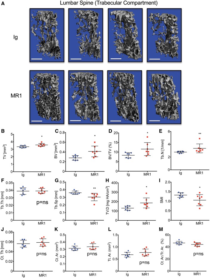
Fig. 2.
MR1 promotes trabecular bone accretion in lumbar vertebrae of skeletally mature mice
Mice were administered Ig (control) or MR1 for 6 months, beginning at 5 months of age and L3 lumbar vertebrae analysed by micro-CT (μCT). (A) Representative μCT reconstructions of trabecular bone. Four representative images are shown for each group. The white scale bar = 500 µm. Quantitative microarchitectural indices of vertebral bone volume and structure were further determined. (B) Tissue volume (TV), (C) bone volume (BV), (D) BV/TV, (E) Tb. N, (F) Tb.Th, (G) Tb.Sp, (H) TV.D and (I) SMI. Cortical indices: (J) Ct.Th, (K) cortical bone area (Ct.Ar), (L) total cross-sectional area (Tt.Ar) and (M) Ct.Ar/Tt.Ar. *P < 0.05; **P < 0.01; by Mann–Whitney test. n = 7 Ig and 7 MR1 mice/group. Mean (s.d.). BV/TV: bone volume fraction; Ct.Th: average cortical thickness; Ct.Ar/Tt.Ar: cortical area fraction; SMI: structure model index; Tb.N: trabecular number; Tb.Th: trabecular thickness; Tb.Sp: trabecular separation; TV.D: trabecular volumetric bone density.

