
Figure 1. Plg-RKT null mice cannot successfully lactate.
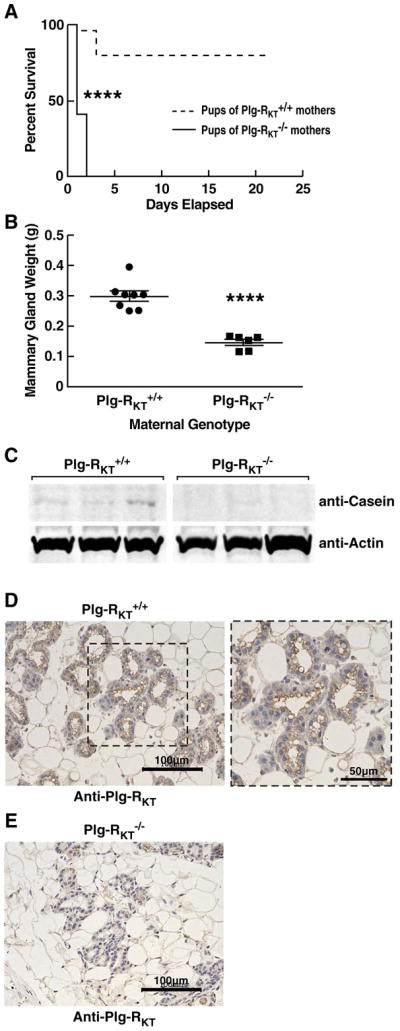
(A), Survival data are shown for a cohort of offspring of Plg-RKT+/+ (dashed lines) and Plg-RKT−/− (solid lines) primiparous female littermates: 48 offspring of Plg-RKT+/+ mice and 30 offspring of Plg-RKT−/− mice. **** P<0.0001 Log-rank (Mantel Cox) test]. (B), Abdominal mammary glands were collected 2 days postpartum from primiparous Plg-RKT+/+ and Plg-RKT−/− female littermates and weighed. **** P<0.0001, n=6 Plg-RKT−/− mice and n=8 Plg-RKT mice+/+ (C), Thoracic mammary glands (harvested 2 days postpartum) were lysed and western blotted with anti-casein and with anti-actin, as a loading control. When using anti-casein antibody, membranes were blocked with LI-COR Casein blocking Buffer. (D,E), Abdominal mammary glands were collected 2 days postpartum from primiparous Plg-RKT+/+ (D) and Plg-RKT−/− (E) female littermates and immunostained with rat anti-Plg-RKT mAb. Images were obtained with a Keyence BZ-X700 (magnification X 200). For the expanded area in panel D, (magnification X 400).