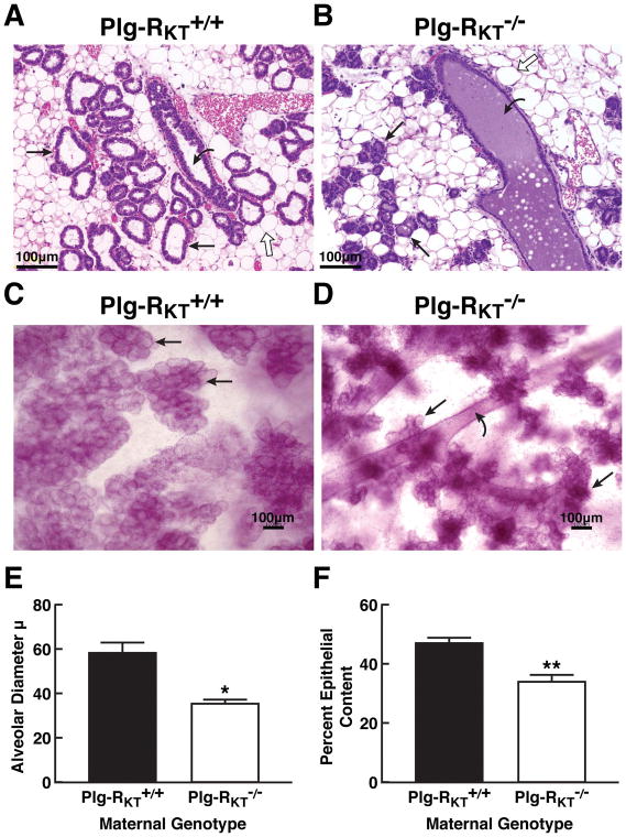
Figure 2. Blockade of lobuloalveolar development in Plg-RKT null mice.
Histology of abdominal mammary glands of Plg-RKT+/+ mice (A) and Plg-RKT−/− littermates (B) harvested 2 days postpartum and stained with H&E. Slides are representative of 6 mice from each group. Images were obtained with a Keyence BZ9000 (magnification X 200). Whole mounts of abdominal mammary glands from the same set of Plg-RKT+/+ mice (C) and Plg-RKT−/− mice (D) harvested 2 days postpartum were prepared as described [39] and stained with carmine red. Images were obtained with a Keyence BZ-X700 (magnification X 100). Straight arrows indicate alveoli, curved arrows indicate ducts and open arrows indicate adipocytes. In Panel C, the visibility of ducts is blocked by the extensive alveoli present. (E), Alveolar diameters of abdominal mammary glands harvested 2 days postpartum. *P=0.01, n=5 Plg-RKT+/+ mice and n=3 Plg-RKT−/− mice. (F), Per cent epithelial content of abdominal mammary glands harvested 2 days postpartum. *P=0.0011, n=6 Plg-RKT+/+ mice and n=3 Plg-RKT−/− mice.

