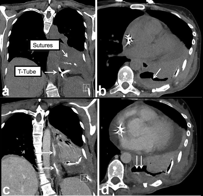
Figure 4.
Nuts and bolts of CT oesophageal leak protocol highlighted using an example. (a) Coronal non-contrast CT image (without oral or intravenous contrast) demonstrates metallic suture material and a T-tube (arrows). (b) Persistent thick-walled basilar loculated hydropneumothorax (arrow) raises the possibility of a leak. (c, d) Coronal and axial images from second phase of the study following administration of oral and intravenous contrast demonstrate extravasation of contrast into the thick-walled collection (double arrows) confirming the leak (oesophagus—single arrows). Teaching point: since true leaks can be small and subtle, the presence of hardware, suture and residual oral contrast from prior imaging studies can confound and mimic a leak on a single phase study.

