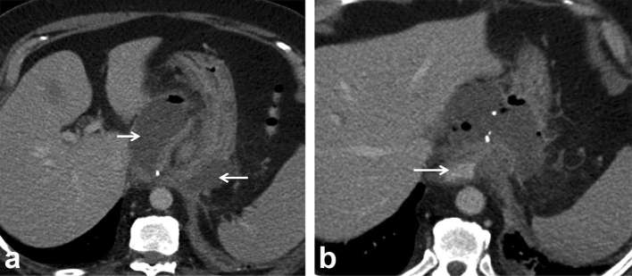
Figure 6.
Nissen fundoplication surgery, fever and peri-anastomotic collection. (a) Axial contrast-enhanced CT image through the upper abdomen shows thick-walled perianastomotic collection (arrows) with fluid and air pockets; suspicious for abscess or infected haematoma vs an underlying leak. (b) Study repeated with oral contrast (arrow) reveals no extravasation, ruling out an oesophageal leak and confirming abscess complicating underlying wrap ischaemia. The patient defervesced with antibiotics and percutaneous drainage. Teaching point: oral contrast helps troubleshoot and characterize the aetiology of peri-anastomotic collections.

