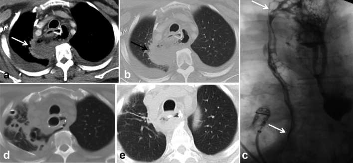
Figure 7.
Post-oesophagectomy, fever and peri-anastomotic collection.(a, b) Axial CT shows thick-walled air and fluid collection adjacent to proximal anastomosis (arrows), extending posterior to the conduit into the right upper lobe. (c) Contrast oesophagogram confirms the presence of a leak extending into the upper lobe (upper arrow) and pleural space (lower arrow). (d, e) Covered oesophageal stent was used to treat leak and subsequent CT shows decreasing size of the collection. Teaching point: mediastinal air/fluid collection may be due to abscess, infected haematoma or ongoing leak. CT with oral contrast or contrast oesophagogram is required to identify underlying anastomotic leak to guide management.

