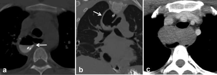
Figure 9.
Post-resection and myomectomy for large bilobed oesophageal leiomyoma. (a, b) Axial and coronal CT images done with oral contrast following surgery show an air-filled mediastinal collection communicating with the oesophagus and filling in with oral contrast (arrows). Since this patient was febrile, this was initially thought to represent a leak at the site of myomectomy. (c) Review of pre-operative CT revealed a large bilobed oesophageal mass distending the oesophagus. The outpouching was correctly interpreted to be redundant oesophagus. Teaching point: differentiating outpouching due to redundant bowel from true leak can be difficult since both fill with oral contrast. Knowledge of the surgical details and pre-operative imaging can help in making the correct diagnosis.

