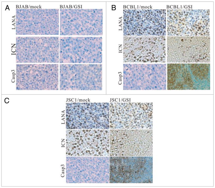
Figure 3.
Immunohistochemical staining of tumor cells. (A) All the signals for LANA, ICN and Caspase 3 proteins in BJAB cells with GSI-treated and mock treated groups were negative. (B) LANA protein in BCBL1 cells with GSI-treated and mock-treated groups were positive. ICN protein in BCBL1 cells with mock-treated groups showed strong positivity, whereas a weak positivity was shown with the GSI-treated group. Caspase3 protein in BCBL1 cells with mock-treated group showed negative stain and was positive in BCBL1 cells from the GSI-treated group. (C) LANA protein in JSC1 cells with GSI-treated and mock-treated groups were positive. ICN protein in JSC1 cells with mock-treated groups showed strong positivity, whereas weak signals positive for ICN was seen in the GSI-treated group. Caspase3 protein in JSC1 cells with mock-treated group showed negative staining and was positive in JSC1 cells with GSI-treated group. (SP ×250).

