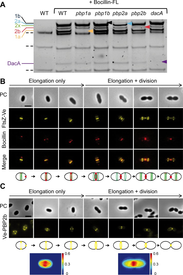
Fig 2. Localization of PBPs and PBP2b during the vegetative cell cycle of L. lactis.
(A) Labelling of L. lactis PBPs by Bocillin-FL staining. Membranes of wild type (WT), pbp1a, pbp1b, pbp2a, pbp2 and dacA mutant cells were purified, incubated with Bocillin-FL (+ Bocillin-FL), and separated on SDS polyacrylamide gel. Bocillin-FL-labeled PBP bands were revealed by fluorescence scanning. Dotted lines indicate auto-fluorescent bands detected in wild-type extracts prior to Bocillin-FL staining. Colored arrowheads mark the absence of PBP1b (1b, black), PBP2a (2a, light blue), PBP2b (2b, red), PBP1a (1a, yellow) and DacA (purple) in the mutant profiles, except for PBP2x whose deleted mutant is not viable. (B) Localization of PBPs with respect to FtsZ during the cell cycle. Cells expressing FtsZ-Ve (NZ3900 [pGIBLD031]) were stained with Bocillin™650/665 and visualized by phase contrast (PC) and epifluorescence (FtsZ-Ve and Bocillin) microscopy. Merge shows the superimposition of both fluorescent patterns. Scale bar, 2μm. L. lactis cell cycle was reconstituted from individual cells taking the beginning of cell constriction (as visualized by phase contrast) as the demarcation between elongation-only and combined elongation + division. PBP staining by Bocillin™650/665 is depicted in red on the cell cycle diagram shown below the pictures. Scale bar, 2μm. (C) Cells expressing the Venus-PBP2b fusion (Ve-PBP2b) (NZ3900 [pGIBLD041]) were visualized by phase contrast (PC) and epifluorescence microscopy. Scale bar, 2μm. A complete cell cycle was reconstituted from representative cells as reported in panel B. Ve-PBP2b fluorescence pattern is depicted in yellow on the cell cycle diagram shown below the pictures. The two bottom panels show fluorescence intensity maps (in arbitrary units, A.U.) from low (blue) to high (red) intensity). Cells (n = 20) were chosen before and after cell constriction based on phase contrast imaging and their normalized Ve-PBP2b fluorescent profiles were superimposed.

