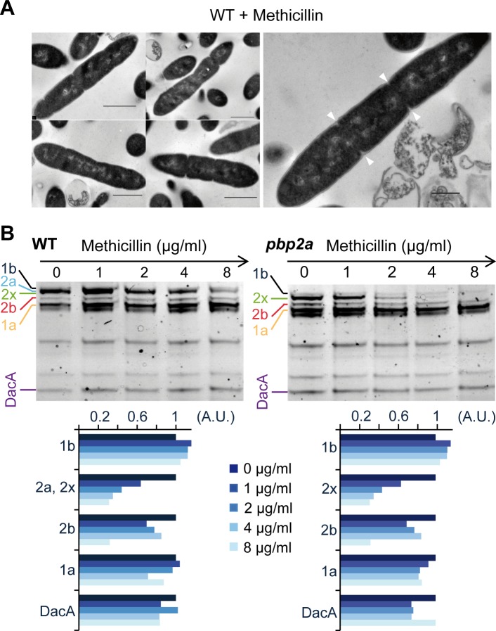
Fig 3. Filamentation of L. lactis induced by methicillin treatment.
(A) Micrographs of wild-type (NZ3900) cells treated by methicillin (1 μg ml-1) obtained by transmission electron microscopy (TEM). Arrows indicate incomplete septa. Scale bars, 500 nm. (B) Identification of methicillin-targeted PBPs by Bocillin-FL staining competition assay. Membrane extracts from wild-type and pbp2a mutant cells were incubated with 0, 1, 2, 4 or 8 μg ml-1 of methicillin prior to add Bocillin-FL. Note the sharp decrease in PBP2x band intensity in the profile of the pbp2a mutant. In wild-type extracts, selective blocking of PBP2x by methicillin is masked by the co-migrating PBP2a band. The two bottom panels show the relative fluorescence intensity of each band (in arbitrary units, A.U.) normalized to the fluorescence intensity measured in absence of methicillin (first lane of each gel).

