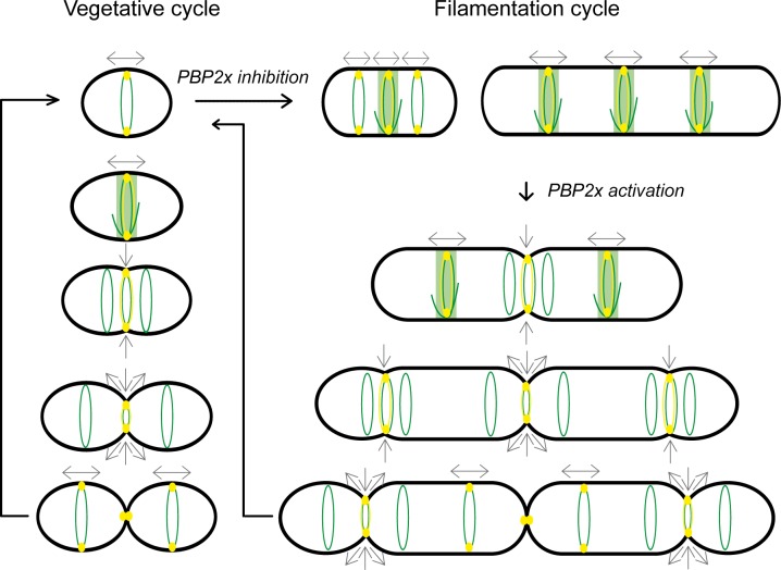
Fig 9. Model for FtsZ-directed dynamics of PBP2b during vegetative and filamentation cycles of L. lactis.
FtsZ structures are shown in green. PBP2b is depicted as a yellow oval. Peripheral localization of the proteins is shown as a ring of the corresponding color. Direction of peripheral and septal growth is shown by grey arrows. The vegetative cell cycle (left) is divided in two separate phases as determined by FtsZ rings structural changes and spatio-temporal localization of PBPs, including PBP2b. During the elongation-only phase (two top cells), FtsZ equatorial ring exhibits a dynamic structure and the PBP2b-dependent peripheral growth mediates cell elongation at mid-cell. At the time of cell constriction, the Z ring segregates into 3 discrete rings; a central constricting ring directing cell division and two lateral rings that move apart as peripheral growth continues from PBP2b located at the septum. After completion of cell division, the elongation-specific PBP2b relocates to the new equatorial FtsZ rings of the newborn cells to reinitiate the cell cycle. The methicillin-induced filamentation cycle (right) results from PBP2x inhibition and reactivation following methicillin removal. During filamentation, PBP2b-dependent peripheral growth mediates cell elongation from a pre-septal position. During filament reversion, FtsZ-directed dynamics of PBP2b takes place as observed during the vegetative cycle but in a highly hierarchical manner starting from the center of the filament.

