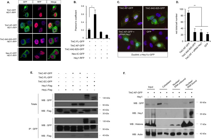
Fig 2. TrkC-KF associates to the transcription factor Hey1 in the nucleus, and both bind to the chromatin.
(A,B) Expression of TrkC-KF-GFP and Hey1-RFP in N2A cells indicates a partial colocalization in the nucleus, as shown by confocal analysis (A) and by the associated Pearson’s coefficient (B). As controls, TrkC, TrkC-642-825, and Neo-IC (fused to GFP) were also transfected. Data represent mean ± SEM (2 independent experiments). *p < 0.05. t test compared to control (TrkC-GFP). (C) Proximity ligation assay (DuoLink) using an anti-Hey1 antibody (recognizing endogenous Hey1) and an anti-GFP antibody on SHEP cells transfected with TrkC-KF-GFP, TrkC-642-825-GFP, GFP, or TrkC-KF-GFP and an siRNA against Hey1: The protein–protein interactions are visualized by red fluorescent spots (Cy3). (D) Quantification of the proximity ligation assay presented in (C): Data represent mean ± SEM (4 independent fields). **p < 0.01. t test compared with TrkC-KF. (E) Immunoprecipitation of TrkC-KF-GFP and TrkC-642-825-GFP using an anti-GFP antibody in HEK293T transfected cells. Hey1 and HeyL are revealed by an anti-Flag WB. (F) HEK293T cells transfected with TrkC-KF-GFP and Hey1-Flag constructs were fractionated into cytoplasmic, nuclear/DNA-unbound, and nuclear/DNA-bound fractions. Actin and Histone H3 are used as loading controls. “Input” corresponds to construct expression in whole cell lysates. Underlying data can be found in S1 Data. GFP, green fluorescent protein; HEK293T, human embryonic kidney 293 T; IP, immunoprecipitation; N2A, Neruo2a; Neo-IC, intracellular fragment of Neogenin; RFP, red fluorescent protein; siRNA, small interfering RNA; TrkC, tropomyosin receptor kinase C; TrkC-FL, full-length TrkC; TrkC-KF, TrkC killer-fragment; WB, western blot.

