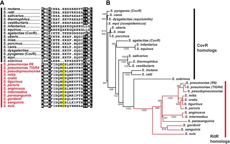
Fig 5. Conservation of RitR in the streptococci.
(A) Alignment of linker regions of RitR homologs (in red) and CovR homologs (in black) from the streptococci. Notice the degeneracy in the “HCS” motif in the swine zoonotic pathogen S. suis. Identical residues are colored black and similar residues are colored grey. The conserved cysteine is shaded in yellow. (B) Phylogenetic tree of RitR (in red) and CovR (in black) streptococcal homologs. Evolutionary distance is depicted by the length of the horizontal lines. Posterior probabilities are displayed at the branch points.

