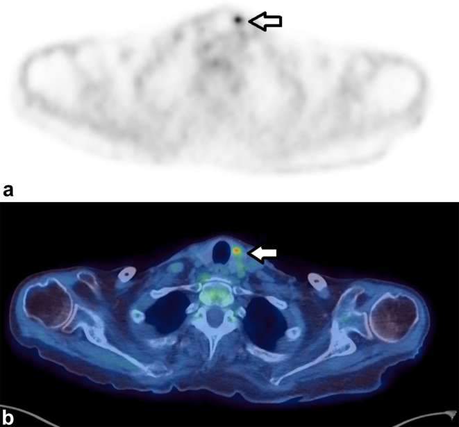
Figure 2.
Axial images of a 71-year-old female who had a PET/CT to assess for possible malignancy as a paraneoplastic cause of her necrotizing autoimmune myopathy. A focus of increased uptake (SUVmax 6.1) (a) localized to the thyroid gland (b) Fine-needle aspiration showed blood and clumps of colloid and clusters of crowded epithelial cells with enlarged nuclei, the appearances were suspicious of papillary carcinoma. Although the study was performed to look for malignancy, this finding was not thought to be the cause of her myopathy. PET, positron emission tomography; SUV, standardized uptake value.

