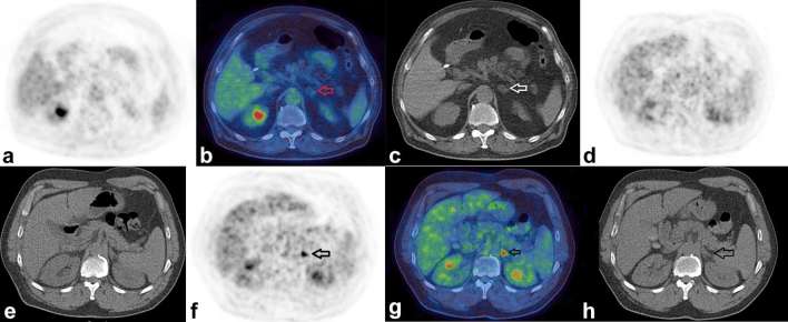
Figure 7.
Benign and malignant adrenal nodules. Figure shows the PET-only (a) fused (b) and CT-only (c) images respectively of a 72-year-old male who had a PET/CT to assess for possible large vessel vasculitis. There was no evidence for vasculitis, however there was a 1.5 cm low attenuation nodule (HU –30) in the left adrenal demonstrating only low-grade FDG uptake (SUVmax 1.5). Based on the presence of macroscopic fat, this was presumed to be an adrenal myelolipoma. Figures (d, e) show PET- and CT-only images respectively in a 68-year-old male who had a PET/CT to further characterize persistent consolidation in his lung, the adrenals look normal. A PET/CT repeated 9 months later (f, g, h) now shows an intense focus of FDG uptake in the left adrenal gland with SUVmax 7.3 (SUVmax of liver 4.1, HU of the adrenal 28). This was assumed to be an adrenal metastasis from his lung cancer which had also progressed elsewhere. FDG, fluorodeoxyglucose; HU, Hounsfield unit; PET, positron emission tomography; SUV, standardized uptake value.

