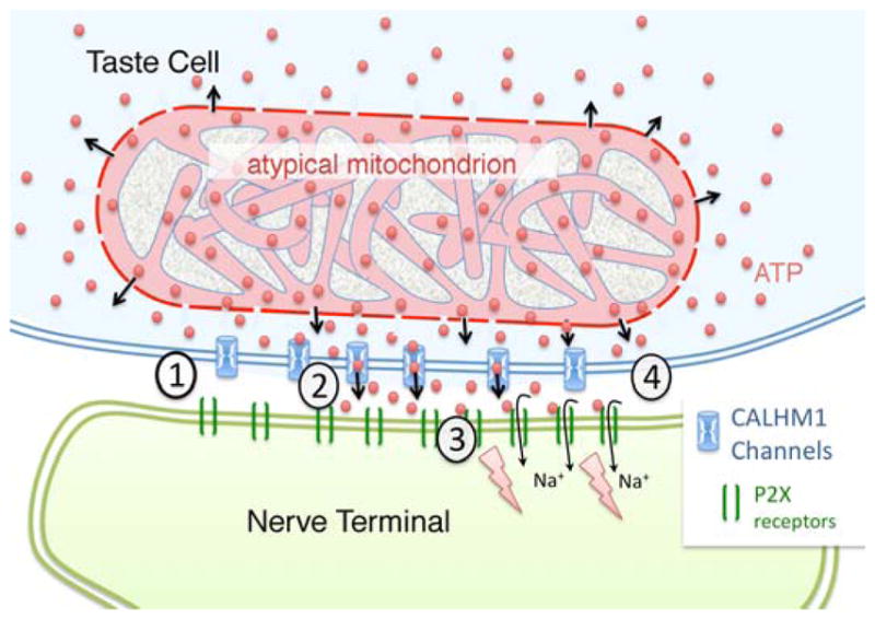
Fig. 7. Schematic diagram of a mitochondrial/CALHM1 synapse between a Type II taste cell (pale blue) and an afferent nerve terminal (green).

The CALHM1 channels lie within the plasma membrane of the taste cell between the atypical mitochondrion (pink) and the nerve terminal. In the resting state (1), CAHLM1 channels (Blue) are closed and ATP levels in the presynaptic compartment will be close to levels in the intermembrane space of the mitochondrion due to diffusion of ATP (red balls) through the porin channels of the outer mitochondrial membrane. Taste evoked action potentials in the taste cell open the CALHM1 channels (2) allowing ATP to flow into the synaptic cleft between the taste cell and the afferent nerve terminal. (3) The ATP in the intercellular space binds to and gates open the P2X receptors (green bars) in the neural membrane, permitting influx of sodium ions (Na+) which depolarizes the afferent terminal. (4) As the taste cell repolarizes, CALHM1 channels close and extracellular levels of ATP return to pre-stimulus levels.
