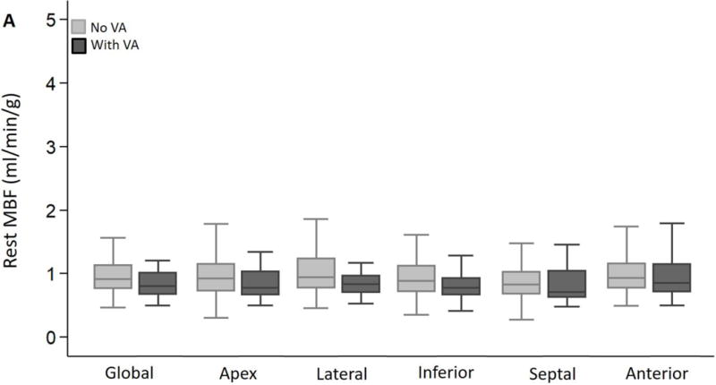
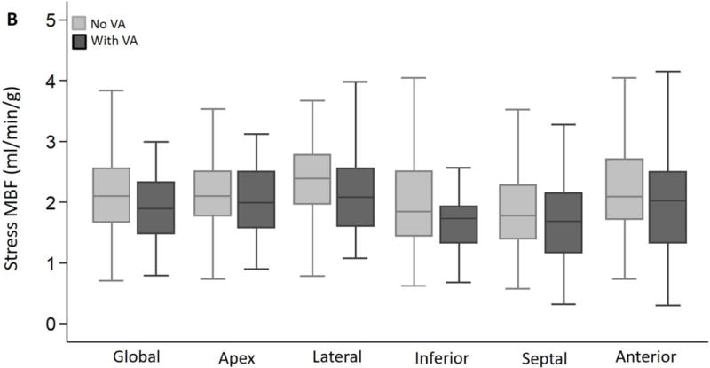
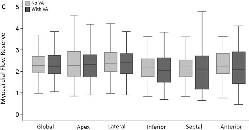
Figure 2. Global and regional MBF and MFR in HC patients stratified by presence or absence of ventricular arrhythmias (VA) during follow up.


(A) Global and regional MBF at rest, (B) Global and regional MBF during vasodilator stress and (C) Global and regional MFR, were similar between HC patients who developed ventricular arrhythmias (sustained VT, NSVT) and HC patients who had no evidence of ventricular arrhythmias during follow up.
