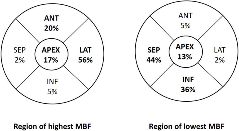
Figure 3. Regional distribution of stress MBF in HC cohort.

The lateral wall demonstrated the highest stress MBF in 56% of patients, and was followed by the anterior wall and apex, respectively, whereas the septum exhibited the lowest stress MBF in ~44% of patients.
