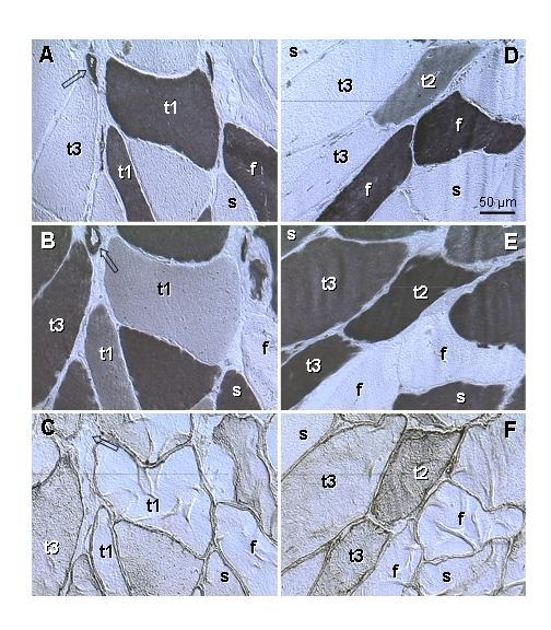
Figure 5.

Type II (A, D) and type I staining (B, E) and p27 expression (C, F) in levator ani muscle of a symptomatic patient (age 50). A-C) Parallel sections. Fast twitch fibers (f) show type II staining only; slow twitch fibers (s) show type I staining only. Some fibers (t1) show strong type II but also weak to moderate type I staining, without cytoplasmic p27 expression. Other fibers (t3) exhibit no type II, strong type I and moderate p27 staining. D-F) Parallel sections. Another type of fibers (t2) shows moderate type II and strong type I staining accompanied by moderate p27 expression, t1-t3 indicates a presumptive course of the type II to type I transition. Arrows indicate vessels, which show both type II and type I staining and no p27 expression.
