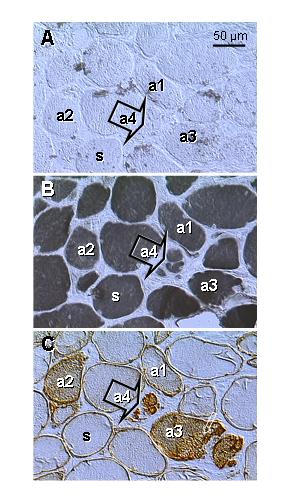
Figure 6.

Type II (A) and type I staining (B) and p27 expression (C) in levator ani muscle of a symptomatic patient (age 63; see also Fig. 3E). Parallel sections. A) No fast twitch fibers are evident. B) All fibers show type I staining, including those exhibiting gradual diminution in size (a3 and a4). C) Cytoplasmic p27 expression gradually increases in regressing fibers (a1 to a4). a1 to a4 indicates presumptive course of apoptosis. s, genuine slow twitch fibers.
