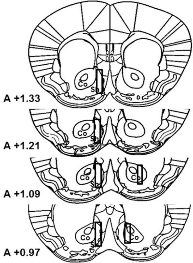
Figure 4.

Drawings of the forebrain sections based on Paxinos and Franklin (2001) with superimposed rectangles that show the confines within which the microdialysis probe tracks were considered to be in the nucleus accumbens shell or core. Data were only included from all subjects with probe tracts within the rectangles. The anterior coordinate (measured from the bregma) is located on each section.
