Figure 1.
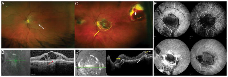
Exudative age related macular degeneration (AMD) with a disciform scar covering the macular region (A). OCT shows cystic intraretinal fluid overlying the subretinal hyperreflective material with loss of the outer retinal layers (B red arrow).
Six months after surgery, the retinal pigment epithelium (RPE)-choroid graft is visualized in the center of the macular region (C, yellow arrow) and the RPE-choroid graft temporal harvest site is flat (white asterisk). The peripheral superotemporal harvesting site of the retinal graft is not visible in the picture.
Postoperative OCT (D) shows the RPE-choroid graft, with rounded hyporeflective spaces due to dilatation of choroidal vessels inside the RPE-choroid graft (white arrows). The neurosensory retinal graft appears well integrated between the native macula, and the RPE-choroid graft separated by a hyper-reflective line (yellow arrows). The retinal layers are not well recognizable. There is resolution of the intraretinal fluid. The silicone oil reflex is visible. Postoperative fluorescein angiography (FA) and Indocyanine Green (ICG) Angiography images (E) showing the complete revascularization of the choroidal graft. Dynamic ICG angiography revealed choroidal vessels starting at the margin of the graft. Feeder vessels of the choroidal patch seemed to grow in sites of contacts of the choroidal graft with healthy and original underlying choroidal vasculature (yellow arrow).

