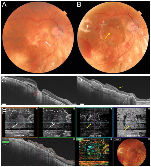
Figure 2.
Patient with advanced exudative AMD (A) with RPE atrophy in the macular region (white arrow). OCT (C) showing lamellar macular hole configuration with a very thin residual foveal floor, significant loss of outer retinal layers and RPE atrophy (red arrow), overlying intraretinal fluid and subretinal hyporeflective spaces. Postoperative photograph 1 month after surgery (B) showing the choroidal-RPE graft in the center of the macular region and the neurosensory retinal graft covering the macular hole (yellow arrow). The temporal harvest site of the neurosensory retinal graft is shown with the white asterisk. Postoperative OCT 3 months after surgery (D) showing RPE-choroidal patch in the macula area, with rounded hyporeflective round spaces due to dilatation of choroidal vessels inside the graft (white arrows). The neurosensory retinal graft (yellow arrow) appears well integrated into the macular hole and is partly lying over the original retina, separated by a hyper-reflective line. Postoperative OCT angiography (E, scan area 6 x 6 mm) 3 months after surgery. The choroidal graft (yellow arrows) is visualized on the en face images at the level of the outer retina and choriocapillaris. The superficial and deep retinal vasculature appear grossly preserved in the area of the original overlying neurosensory retina (white arrows), however the scan is limited by artifact and segmentation errors.

