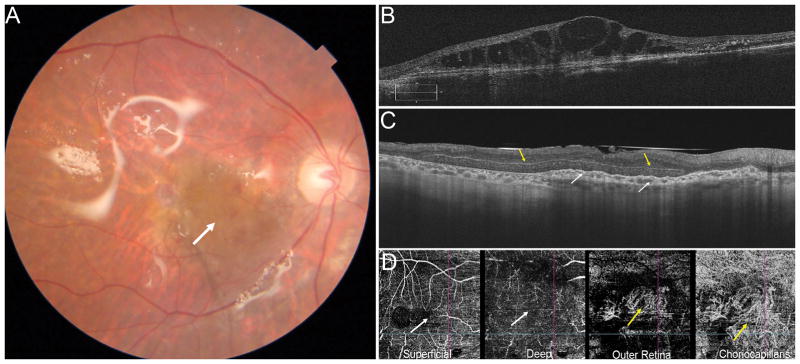
Figure 4.
Postoperative fundus photograph showing the RPE-choroidal free graft in the center of the macula (A. white arrow). The neurosensory retinal free graft is under the native retina. The temporal harvest site of RPE-choroidal patch and the superotemporal harvest site of the retinal patch are not visible. Preoperative OCT showing advanced exudative AMD with subretinal hyperreflective material, loss of outer retinal layers and overlying cystic intraretinal fluid (B). Postoperative OCT (C) showing the RPE-choroid graft in the macula area, with a dome-shaped aspect due to dilatation of choroidal vessels and an irregular surface secondary to contraction of the RPE-choroid graft (white arrows). The neurosensory retinal free graft is well integrated between the native retina and the RPE-choroidal graft, separated by a hyper-reflective line, (yellow arrow) and there is relative preservative of the retinal architecture. OCT angiography (D, scan area 6 x 6 mm) shows the area of the RPE-Choroid graft seen on the outer retina and choriocapillaris slabs (yellow arrows). While the superficial capillary plexus is relatively well preserved (left white arrow), the deep capillary plexus which is likely in the area of the neurosensory graft (right white arrow) is not well visualized. The scan is however limited by artifact and segmentation errors.

