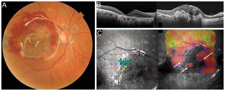
Figure 5.
Fundus photograph after surgery (A) showing the RPE-choroid free graft in the center of the macular region with the overlying neurosensory retinal graft (white arrow). The harvest sites of the RPE-choroid free graft temporally and the neurosensory retinal free graft superotemporally are not visible in the picture. Preoperative OCT (B, left) showing an atrophic macular area with loss of the outer retinal layers and highly reflective subretinal material (red arrow). Postoperative OCT (B, right) showing the RPE-choroid graft with dilatation of choroidal vessels and an irregular surface (white arrows). The choroidal patch is vascularized as shown by the ovoidal dark hyporeflective spaces. The intraretinal neurosensory retinal free graft appears well integrated (yellow arrow) with some inner retinal boundaries visible overlying the RPE-choroidal free graft. Postoperative microperimety (C) and fixation maps (4-2 strategy). The area of fixation was at superotemporal border of the retinal free graft (yellow arrow) overlying the area of the choroidal graft. There was a dense scotoma with complete loss of sensitivity, however there are areas of low sensitivity along on the retinal free graft, in the superotemporal, nasal and temporal sides and over the choroidal graft (white arrows).

