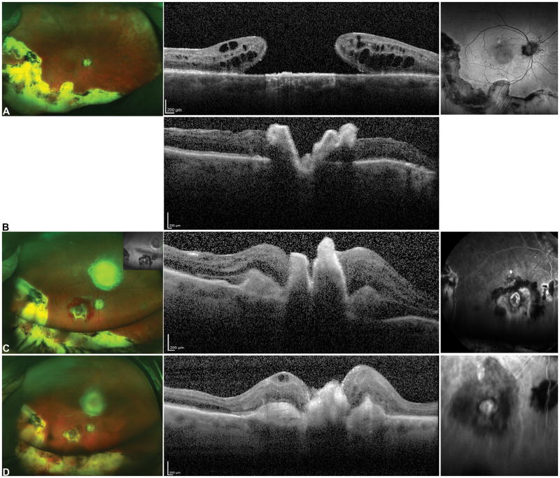
Figure 7.
76-year-old M with exudative age related macular degeneration with a concurrent large macular hole refractory to prior internal limiting membrane removal and chorioretinal scarring from prior retinal detachment repair. Preoperative OCT (A, middle) shows a large refractory macular hole with 2400-micron largest basal diameter and 1600-micron inner opening diameter. Following a combined autologous neurosensory retinal and choroidal free graft with silicone oil tamponade (B), post-operative day 1 OCT (C) shows the choroidal graft plugging the macular hole, the neurosensory retinal graft had dislocated. Postoperative photograph at 1 month shows the choroidal free graft covering the macular hole, free graft harvest site and a silicone oil tamponade. OCT at 2 months (C) and shows improved integrated of the choroidal free graft tissue in the macular hole with surrounding retinal tissue. Fluorescein angiogram (C, right) shows blockage from the subretinal heme, staining around the choroidal graft site and no leakage. The subretinal hemorrhage resolved. OCT at 12 months shows resolution of subretinal fluid and further improved integration of the choroidal free graft with the retinal tissue and closure of the macular hole and ICG shows blockage in the area of the choroidal graft with no abnormal vascular complex or leakage. The vision was improved from HM preoperative to 20/200E@1 meter 12 months postoperatively with subjective improvement as well.

