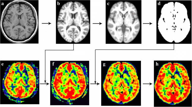
Fig. 1.
Methods of CBF normalization and standardization. The individual raw T1 images (a) were segmented and generated T1 tissue probability maps (b), which would be used to generate average T1 tissue probability maps (c) and brain mask (d). The individual CBF maps (e) was coregistered by the T1 tissue probability maps, which would generate normalized CBF maps (f). The brain mask was applied with the normalized CBF maps in order to extract the brain tissue (g), and the Z transformation was performed with the normalized CBF maps to standardize the CBF maps (h)

