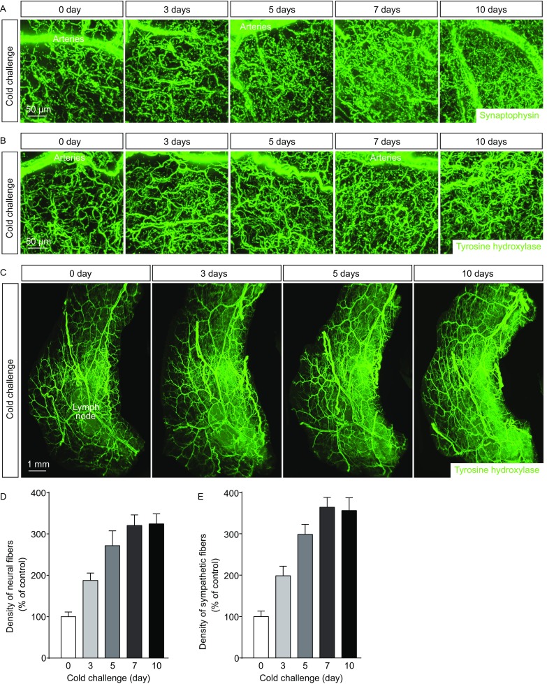
Figure 1.

Intra-adipose sympathetic plasticity in response to cold challenge. (A–E) The wildtype mice were subject to cold challenge. (A) Representative 3D projections of iWAT harvested at indicated time points, immunolabeled by anti-synaptophysin, and imaged at 12.6× magnification on the lightsheet microscope. (B and C) Representative 3D projections of iWAT harvested at indicated time points, immunolabeled by anti-tyrosine hydroxylase, and imaged at 12.6× (B) or 1.26× (C) magnification on the lightsheet microscope. (D) Density of the neural fibers in iWAT immunolabeled by anti-synaptophysin was quantified. n = 4, mean ± SEM. (E) Density of the sympathetic fibers in iWAT immunolabeled by anti-tyrosine hydroxylase was quantified. n = 5, mean ± SEM
