
Figure 5.
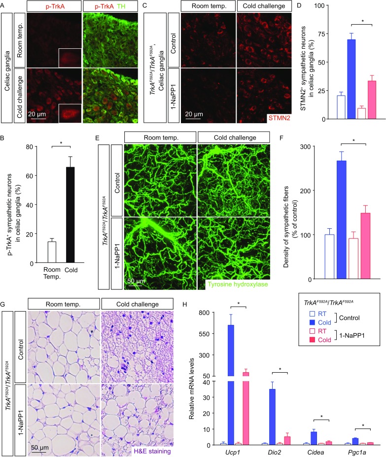
Intra-adipose sympathetic plasticity regulated by the TrkA signal is required for cold-induced beiging process. (A and B) The wildtype mice were subject to cold challenge. (A) Activation of the TrkA signal in sympathetic neurons of the celiac ganglia was examined by conventional immunostaining of anti-p-TrkA. Insets, representative zoom-in views of vesicular p-TrkA localization. (B) p-TrkA-positive sympathetic neurons of the celiac ganglia were quantified. n = 4, mean ± SEM, *P < 0.01. (C–H) TrkAF592A/F592A mice daily-treated with 1-NaPP1 or vehicle control were subject to cold challenge. (C) STMN2 expression in sympathetic neurons of the celiac ganglia was examined by conventional immunostaining. (D) STMN2-positive sympathetic neurons of the celiac ganglia were quantified. n = 4, mean ± SEM, *P < 0.01. (E) iWAT was processed for the volume fluorescence-imaging of anti-tyrosine hydroxylase. Representative 3D projections of iWAT imaged at 12.6× magnification on the lightsheet microscope. (F) Density of the sympathetic fibers in iWAT was quantified. n = 5, mean ± SEM, *P < 0.01. (G) Appearance of multilocular beige cells in iWAT was examined by H&E staining. (H) Expression levels of the beiging-related genes in iWAT were determined by the qPCR analysis. n = 5, mean ± SEM, *P < 0.01