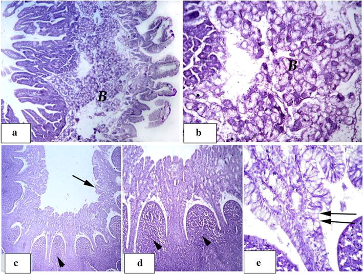
Fig. 4.

Group T5: a showing significant development in duodenum wall. b Showing massive layer of mixed brunners gland filling almost of the submucosa. c–e normal, regular and intact lymphatic nodules; peyers patches within the ileum submucosa (arrow head), intact villi (arrow) with increase in goblet lining cells (double arrows)
