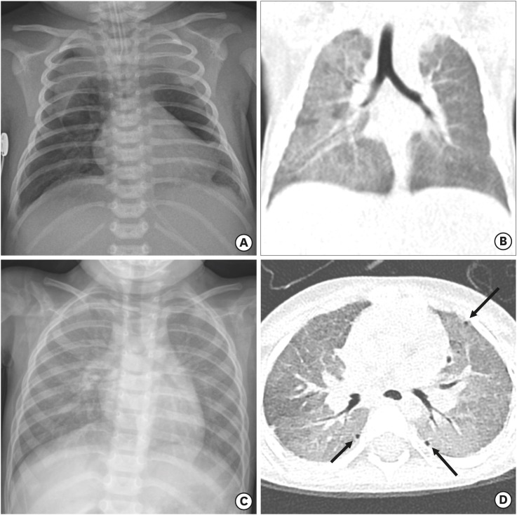
Fig. 1. Sequential chest radiographs and CT. (A) Chest radiograph on the second postnatal day reveals a pneumomediastinum during the treatment with mechanical ventilation. (B) Coronal reconstructed chest CT on postnatal day 18 shows a resolved pneumomediastinum. However, there is residual diffuse GGO in both lungs with the right lung predominance. (C) Chest radiograph at 20 months of age demonstrates recurrence of diffuse haziness in bilateral lungs. (D) Axial chest CT image at 20 months of age shows diffuse GGO with mosaic attenuation and multiple tiny subpleural air cysts (arrows) in both lungs.
CT = computed tomography, GGO = ground-glass opacity.

