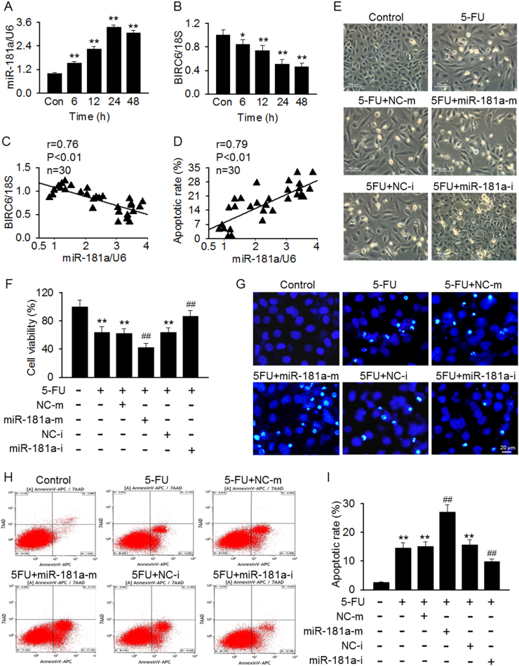
Fig. 1. MiR-181a regulates 5-FU-induced mesangial cell apoptosis.
a, b Analysis of miR-181a (a) and BIRC (b) in mesangial cells treated with 5-FU (100 μM) for 6, 12, 24, or 48 h. **p < 0.01 vs. control, n = 6. c, d miR-181a expression was inversely correlated with BIRC6 mRNA expression (c) and positively correlated with apoptotic rate (d). e The cells were pretreated with miR-181a mimics or miR-181a inhibitor for 48 h followed by incubation of 5-FU (100 μM) for another 24 h. Cell morphology was assessed by phase contrast microscopy. f Cell viability was assessed by CCK-8 assay. **p < 0.01 vs. control; ##p < 0.01 vs. 5-FU, n = 6. g Hoechst 33258 nuclei staining was used to detect apoptotic morphology. h Fluorescence-activated cell sorting-derived dot plot diagrams of mesangial cells stained with annexin V-APC/7-AAD. i Percentage of apoptotic cells was determined by quantitative analysis. **p < 0.01 vs. control;##p < 0.01 vs. 5-FU, n = 4

