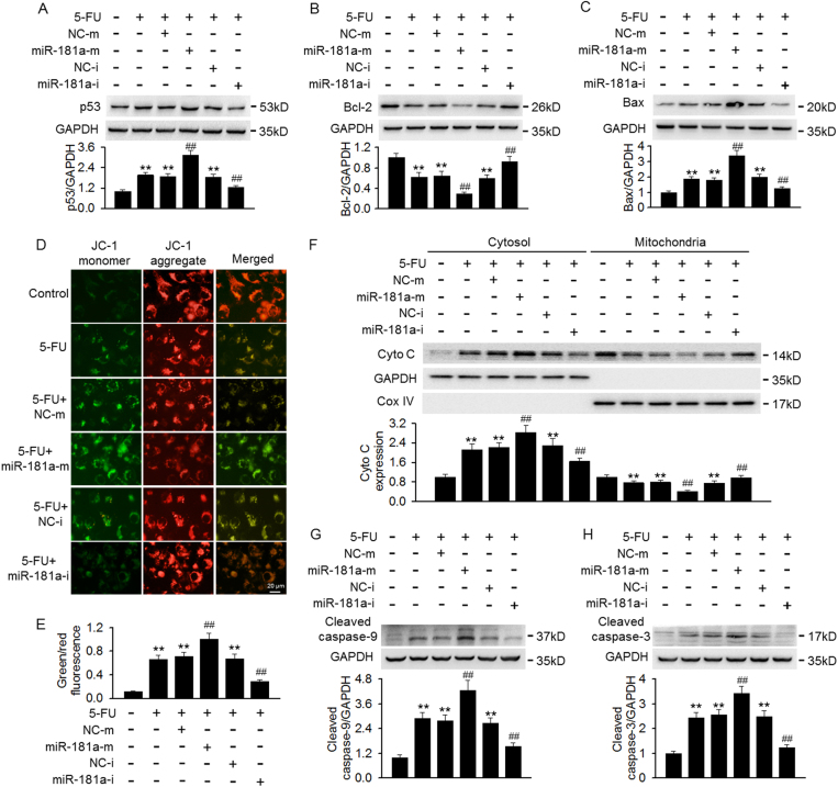
Fig. 2. Effects of miR-181a on p53-dependent apoptosis signaling after 5-FU treatment.
a–c Cells were pretreated with miR-181a mimics or miR-181a inhibitor for 48 h prior to incubation of 5-FU (100 μM) for another 24 h. The protein expression of p53 (a), Bcl-2 (b), and Bax (c) were determined by western blotting. d Mitochondrial membrane potential was measured by JC-1 staining. e Quantitative analysis of the green/red fluorescence intensity ratio, which represented as a surrogate marker of mitochondrial membrane depolarization. f Western blotting analyses of 5-FU-induced mitochondrial cytochrome c (Cyto c) release from mitochondria into the cytosol. g, h Caspase-9 (g) and caspase-3 (h) cleavage were detected by western blotting. **p < 0.01 vs. control; ##p < 0.01 vs. 5-FU, n = 6

