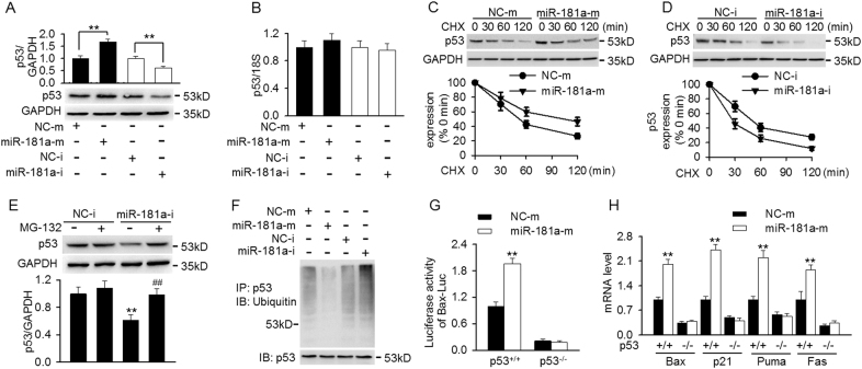
Fig. 3. MiR-181a inhibition decreased p53 protein expression by facilitating ubiquitin-mediated degradation.
a, b Cells were transfected with miR-181a mimics or miR-181a inhibitor for 48 h. The protein (a) and mRNA (b) expression of p53 were determined by western blotting and qRT-PCR, respectively. **p < 0.01 vs. miRNA negative control or miRNA inhibitor negative control, n = 5. c, d Mesangial cells pretreated with miR-181a mimics (c) or miR-181a inhibitor (d) were incubated with cycloheximide (CHX, 10 μg/ml) for indicated time period. Representative western blotting image of p53 and relative intensity of p53 protein expression were shown. e Cells were treated with or without MG-132 (20 μM) for 12 h prior to transfection with miRNA inhibitor negative control or miR-181a inhibitor for another 48 h. P53 protein expression was analyzed by western blotting. **p < 0.01 vs. miRNA inhibitor negative control; ##p < 0.01 vs. miR-181a inhibitor, n = 6. f Cell lysates were immunoprecipitated with p53 antibody and the ubiquitination of p53 was detected by western blotting using ubiquitin antibody. g HCT116 p53+/+ cells and HCT116 p53−/− cells were co-transfected with miR-181a mimics or negative control and Bax luciferase (Bax-Luc) which contains the binding site of p53 for luciferase activity assay. h The mRNA expression of p53 target genes, including Bax, p21, Puma and Fas, was detected in HCT116 p53+/+ cells and HCT116 p53−/− cells after miR-181a mimics transfection. **p < 0.01 vs. miRNA negative control, n = 6

