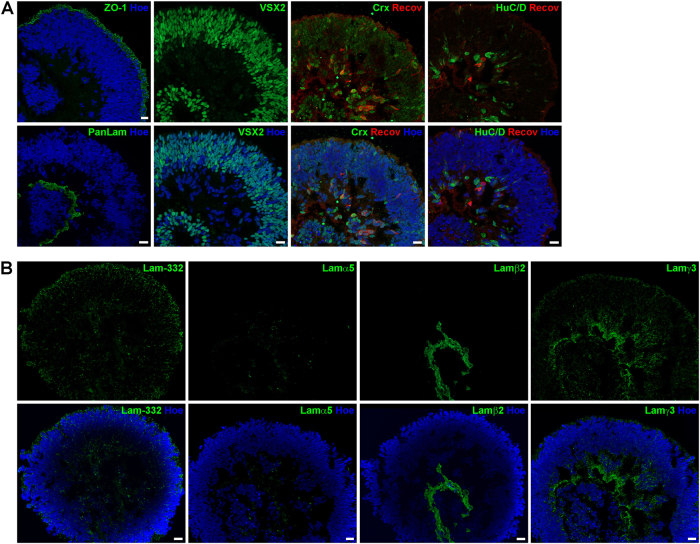
Fig. 3. Expression of retinal markers and laminin-332, laminin α5, laminin β2 and laminin γ3 in retinal organoids derived from hESCs at day 35 of differentiation.
a) Expression of ZO-1 (green), Pan-Laminin (PanLam, green), retinal progenitor cells (VSX2, green), photoreceptors (CRX, green; Recoverin, red) and ganglion cells (HuC/D, green). b) Laminin-332 (green) is expressed throughout the retinal organoid. No immunoreactivity for laminin α5 (green) was found. Laminin β2 (green) was observed in a basement membrane-like structure at the basal site of the retinal organoid. Laminin γ3 (green) is expressed throughout the retinal organoid with a prominent labelling at the basal site. Nuclei are counterstained with Hoechst (blue). Hoe Hoechst, Lam laminin, PanLam Pan-Laminin, Recov Recoverin. Scale bars, 20 μm

