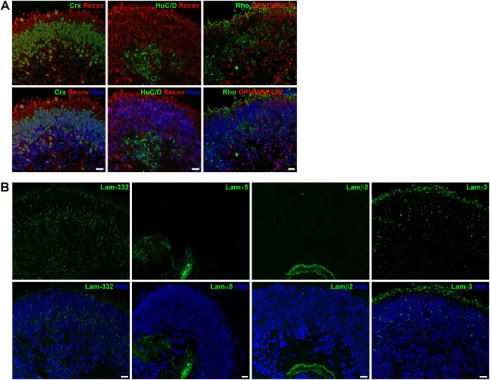
Fig. 6. Expression of retinal markers and laminin-332, laminin α5, laminin β2 and laminin γ3 in retinal organoids derived from hESCs at day 200 of differentiation.
a) Expression of photoreceptors (CRX, green; Recoverin, red), ganglion cells (HuC/D, green), rods (Rho, Rhodopsin, green) and cones (OPN1MW/OPN1LW, red). b) Laminin-332 (green) is expressed throughout the retinal organoid with a prominent labelling in the middle of the organoid. Laminin α5 (green) and laminin β2 (green) were observed in a basement membrane-like structure at the basal site of the retinal organoid. Laminin γ3 (green) was found predominantly at the apical site of retinal organoid with a prominent labelling above cell nuclei, most likely in the IS of photoreceptors. Nuclei are counterstained with Hoechst (blue). Hoe Hoechst, IS inner segments, Lam laminin, Recov Recoverin, Rho Rhodopsin. Scale bars, 20 μm

