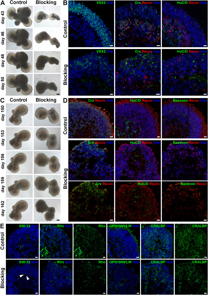
Fig. 7. Blocking of laminin γ3 in retinal organoids derived from hESCs at day 43 and day 150 of differentiation.
a) Brightfield images revealed a degeneration of bright phase retinal neuroepithelial structures at the apical edge of the organoids in the blocking condition over time in the early blocking experiment (day 43). b) Expression of retinal progenitor cells (VSX2, green), photoreceptors (CRX, green; Recoverin, red) and ganglion cells (HuC/D, green) showed a reduction in cell number and a disruption of the lamination in retinal organoids after blocking of laminin γ3 at day 43 of differentiation. c) Brightfield images indicated a degeneration of the retinal neuroepithelial structures at the edge of organoids over time upon blocking of laminin γ3 at day 150 of differentiation. d) Expression of photoreceptors (CRX, green; Recoverin, red) indicated the lamination loss in retinal organoids after blocking of laminin γ3 at day 150 of differentiation. Expression of ganglion cells (HuC/D; green) were absent in the blocking condition. Bassoon (green) expression is significantly decreased in blocked-retinal organoids. e) After laminin γ3 blocking only a few ganglion cell dendrites detected by SMI-32 (green) were still present (arrowheads). Expression of rods (Rho, green), cones (OPN1MW/OPN1LW, green) and Müller cells (CRALBP, green) revealed the disrupted lamination of retinal organoids and a reduction in their cell number at day 150 of differentiation. Nuclei are counterstained with Hoechst (blue). Hoe Hoechst, Recov Recoverin, Rho Rhodopsin. Scale bars, 50 μm for a and c; 20 μm for b, d, e

