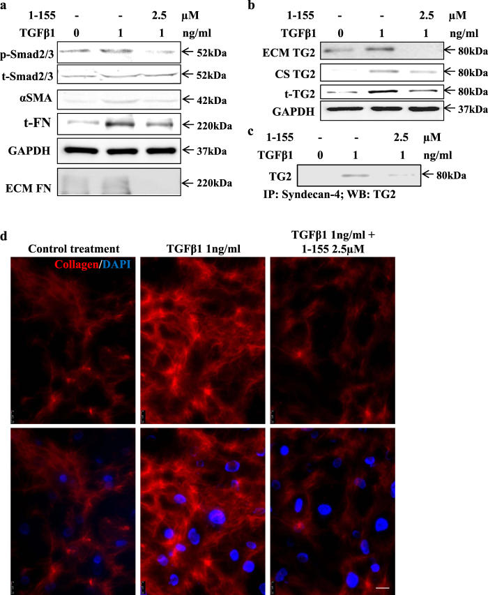
Fig. 4. The effect of TG2 inhibition on TGFβ1-induced cardiofibroblast to myofibroblast transition and collagen deposition in human cardiofibroblasts.
a Representative western blots (n = 3) showing p-Smad2/3 activation, αSMA and FN and FN deposition and the inhibition of these parameters using TG2 inhibitor 1–155 (2.5 µM) in 72 h TGFβ1-treated cardiofibroblasts at the concentrations shown. Matrix FN was measured according to the procedures described in the 'Materials and methods'. b Representative western blot (n = 3) showing the effect of 72 h TGFβ1 treatment in cardiofibroblasts showing increased TG2 expression and increased TG2 at the cell surface and ECM and inhibition of this by TG2 inhibitor 1–155. c Representative western blot (n = 3) of TG2 in the syndecan-4 immunocomplex from co-IP carried out as described in 'Materials and methods'. d Immunofluorescence detection of collagen I in cardiofibroblasts treated with 1 ng/ml TGFβ1 over 5 days with and without the TG2 inhibitor at 2.5 µM undertaken as described in the 'Materials and methods'. Bar = 25 µm

