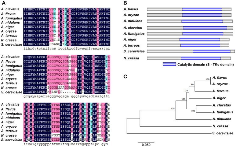
Figure 1.
Bioinformatics analysis of PbsB. (A) Amino acid alignment of A. flavus PbsB and other 8 putative orthologs. Clustal X was used in this analysis. (B) Diagram shows the domains in PbsB among above 9 species. InterPro (http://www.ebi.ac.uk/interpro/scan.html) and IBS 1.0 were used in the analysis. (C) The diagram of the phylogenetic tree among 9 species.

