Fig. 1.

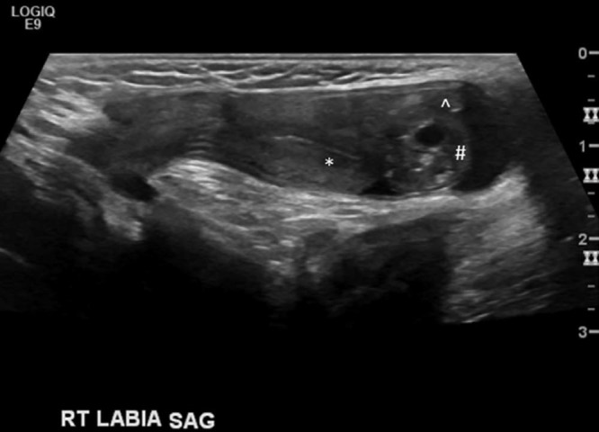
Ultrasound image demonstrating a sagittal view of the right inguinal region shows the canal of Nuck hernia containing the uterus (*), the RT ovary (#), and the fallopian tube (^).

Ultrasound image demonstrating a sagittal view of the right inguinal region shows the canal of Nuck hernia containing the uterus (*), the RT ovary (#), and the fallopian tube (^).